Inferior Vena Cava Filter Placement Care Guide
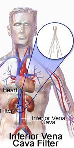 |
Why do I need an IVC filter?
You may need an IVC filter if you have a blood clot in your leg. You may also need an IVC filter if your risk of blood clots is increased, such as after surgery or during pregnancy. You may need a temporary or permanent filter.What happens during IVC filter placement?
Your caregiver will insert a catheter (thin plastic tube) into a blood vessel in your neck or groin. He will use an ultrasound or x-ray to guide the catheter into your IVC. The filter will be pushed through the catheter and attached to the walls of the IVC. The catheter is pulled out and the filter is left in. Your caregiver will press firmly on the area where the catheter went in, to stop any bleeding. After a few minutes, your caregiver will put a bandage on the area.I was awake for the entire procedure. While not terribly painful or even uncomfortable, it was quite odd. I could feel the catheter being inserted and the filter sliding down my vein. While there was very little pain at the time it is VERY disconcerting to feel something going down the inside of your neck.
What are the risks of an IVC filter?
You may bleed more than expected or get an infection. Your IVC and the tissue around it may get damaged during the procedure. Your filter may break, loosen, move, or get blocked. You may need another procedure to fix these problems with your filter.Temporary or retrievable filters (also called optional filters) can usually be retrieved or repositioned up to a certain point in time. With time, the filter becomes incorporated into the caval wall and may not be removable. The Cook Celect and Gunther Tulip filters are optional filters; they have retrieval kits that are used to snare the apical hooks and retrieve the filters. While removal within 30 days is typical, successful filter removal more than 1 year after implantation has been reported
My procedure was done through the neck, the jugular vein (which still hurts by the way)
What does the equipment look like?
In this procedure, a catheter, iodine contrast (x-ray dye), x-ray or ultrasound equipment for imaging guidance and an inferior vena cava (IVC) filter may be used.A catheter is a long, thin plastic tube that is the same size or smaller than a pencil.
X-ray:
The equipment typically used for this
examination consists of a radiographic table, an x-ray tube and a
television-like monitor that is located in the examining room.
Fluoroscopy, which converts x-rays into video images, is used to watch
and guide progress of the procedure. The video is produced by the x-ray
machine and a detector that is suspended over a table on which the
patient lies.
Ultrasound:
Ultrasound scanners consist of a console containing a computer and electronics, a video display screen and a transducer
that is used to do the scanning. The transducer is a small hand-held
device that resembles a microphone, attached to the scanner by a cord.
The transducer sends out inaudible high frequency sound waves into the
body and then listens for the returning echoes from the tissues in the
body. The principles are similar to sonar used by boats and submarines.
The ultrasound image is immediately visible on a video display screen that looks like a computer or television monitor. The image is created based on the amplitude (loudness), frequency (pitch) and time it takes for the ultrasound signal to return from the area of the patient being examined to the transducer, as well as the composition of body tissue through which and the type of body structure the sound travels through.
Other equipment that may be used during the procedure includes an
intravenous line (IV) and equipment that monitors your heart beat and
blood pressure.The ultrasound image is immediately visible on a video display screen that looks like a computer or television monitor. The image is created based on the amplitude (loudness), frequency (pitch) and time it takes for the ultrasound signal to return from the area of the patient being examined to the transducer, as well as the composition of body tissue through which and the type of body structure the sound travels through.
OK, so this was all kind of cool. Even though I had a "tent" over my head it was pulled up enough to offer a view of the screens for the ultrasound and the xray. Watching the die show up and my insides change color was fascinating. As was watching the filter being put in. I could see my intestines, my spine and my ribs. The photos below aren't of my procedure obviously, but they are pretty much exactly what I was able to see.


What are the benefits vs. risks?
Benefits
- No surgical incision is needed—only a small nick in the skin that does not have to be stitched closed.
- The filter has a high rate of success in protecting lungs from serious pulmonary embolus (PE) in patients who have failed conventional medical therapy or cannot be given conventional medical therapy.
Risks
- Any procedure where the skin is penetrated carries a risk of infection. The chance of infection requiring antibiotic treatment appears to be less than one in 1,000.
- There is a very slight risk of an allergic reaction if contrast material is injected.
- Any procedure that involves placement of a catheter inside a blood vessel carries certain risks. These risks include damage to the blood vessel, bruising or bleeding at the puncture site, and infection.
- There is a chance that the IVC filter can lodge in the wrong place, change position or penetrate through the vein (which can rarely lead to injury of a nearby organ).
- The IVC filter or a piece of the IVC filter may break loose and travel to the heart or lungs causing injury or death.
- Rarely, IVC filers become so filled with clots that they block all flow in the blood vessel, causing swelling in the legs.
- In some cases, retrievable filters become scarred to the vein and cannot be removed, in which case they are left in permanently (as they are also designed to do).
No comments:
Post a Comment